

🔬 Biological _ Electrical Nanosensors Section
A complete review of nanobioelectrical nanobiosensors and stripping adsorption voltammetry .
Researcher and Author: Dr. ( Afshin Rashid)
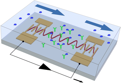
Note: In the construction of electrochemical nanobiosensors, the minimum parts used in a biosensor are: a molecular recognition layer and a signal transducer that can be connected to a readout device for these signals.
DNA is often a suitable tool as a biosensor because the base pairing reaction between complementary base sequences is both specific and stable. In this case, a single-stranded probe DNA is immobilized on a detection layer and then reacts with the probe by the target DNA on the surface through the pairing process. The repetitive nature and uniformity of DNA structures make their aggregation on the surface very specific. It is on this surface that the capture of the target DNA and the generation of the signal occur. Therefore, immobilizing the probe nucleic acid while maintaining its original binding strength is important for the detection of the target DNA. However, how this detection process is measured depends on the method of signal transduction, which may be optical, mechanical, or electrochemical.
Optical biosensors that work based on fluorescence light have the following characteristics. These types of biosensors are sensitive to molecules per square centimeter. They consist of rows that are 7 in number, so that their detection limit is made up of approximately thousands of probes. Because the instruments in this field (fluorescence biosensors) are complex and expensive, gene chip technology is more suitable for laboratory work. Gene chips are more suitable in cases where a lot of work needs to be done simultaneously, such as transcriptional profiling or single nucleotide polymorphism discovery. However, clinical diagnostics usually do not require the collection of this much data. What is important for molecular diagnostics is the reliability of the diagnosis and also the generality regardless of the order of the game. Therefore, gene chips are not preferred for clinical diagnostics for several reasons, such as: they are expensive and the device is complicated, and also for other specific reasons, the accuracy of the reading is reduced. Another method for measuring the signal optically is the surface plasmon resonance method. In this method, a change is created in the refractive index of a thin metal film substrate, which is due to the absorption of the analyte and is suitable for detecting the target in cases that are house-house, groove-groove. In order to be able to reach the detection limit of the target molecule in this method, at which point a signal is generated, the hybridization signal must be This can be enhanced by increasing the amount of material on the film surface before or after binding to the target DNA. Like fluorescence, surface plasmon resonance (SPR) is expensive and complex, which has made it more suitable for research than routine diagnostic work; one of the most specific methods for measuring the signal optically is the optical readout method, in which single-stranded DNA is labeled with gold nanoparticles that change color upon hybridization with the target sequence . Using silver staining, DNA analysis can be performed with this optical method in very small plates with high sensitivity. Although the use of gold nanoparticles may be expensive, this method has the sensitivity and simplicity necessary for clinical diagnostics.
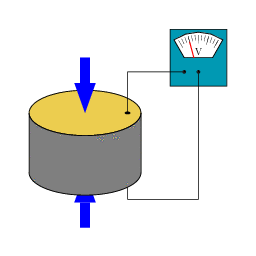
One of the methods of reading or measuring the signal is to measure the changes that occur in the immobilized detection layer due to binding to the target molecule. In this case, a quartz microbalance crystal is most often used. This device is sensitive and can report the production of hybrids (pairing) at the time of their formation. One of the limitations of the QMC method is that this method cannot be performed when the target sample is in the solution phase, but with the advances in this field, namely the creation of new methods of sample amplification and amplification, this limitation may be eliminated. Another method of volumetric measurement is the use of microfabricated cantilevers . In this method, the increase in volume that accompanies hybridization is detected by the deflection of the laser beam from the cantilever surface. The advantages of this method are that it is suitable for developing linear grooves and that non-specific connections can be corrected with this method. The disadvantages of this method are the expensiveness and complexity of the tools and equipment used. Electrochemical methods are well suited for DNA detection because electrochemical reactions directly generate electronic signals, eliminating the need for expensive transducers. Furthermore, because the immobilized base sequence can be confined to a single electrode substrate, detection can be performed by inexpensive electrochemical assays.
Electrochemical sensors are being developed for clinical or environmental testing in situ . The sensitivity of electrochemical signals is based on the direct oxidation or catalysis of DNA bases as well as the reduction reactions of reporter molecules or enzymes. Various methods are used to measure the signal electrochemically. The basis of measuring the signal in the direct DNA electrochemical method is based on the oxidation and reduction reaction of DNA at a mercury electrode, so the amount of oxidized and reduced DNA is proportional to the amount of DNA that hybridizes with the probe. In addition to the old methods of direct DNA reduction, a method called stripping adsorption voltammetry is used today to select the direct DNA oxidation nanomolecules, which is a very sensitive method. In the direct electrochemical method, purine bases are oxidized by materials such as carbon, indium tin oxide (ITO), gold, and polymer-coated electrodes . Although the direct electrochemical method is a very sensitive method, its application is complex.

Conclusion:
All molecular-based electrochemical nanobiosensors rely on a highly specific system to detect or track their target molecule. The importance of an electrochemical nanobiosensor is to provide a suitable support for binding the target molecule to the probe and generating an electrical signal that can be measured and read.
Researcher and Author: Dr. ( Afshin Rashid)
Specialized PhD in Nano-Microelectronics